Stage 01
Consultation
Symptoms heard. History reviewed. The first decision is whether intervention is needed at all.
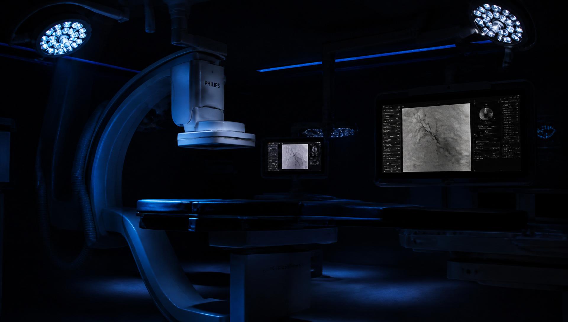
Advanced image-guided treatment through minimally invasive procedures.

Anatomy
From the brain to the feet, explore the blood vessels and the conditions that can affect them.
Stroke · Brain aneurysm · AVM / AVF
Procedures performed
Conditions
Explore conditions affecting the blood vessels of the body, and understand the image guided treatments available for selected cases.
View all conditions →When disease begins inside a vessel, treatment changes.
Image-guided intervention reaches the disease from within, preserving healthy tissue and reducing recovery wherever appropriate.
Procedures
Advanced image-guided procedures are performed through a small access point, with real-time imaging guiding each step.
Procedure 01
Reopening a narrowed artery from the inside.
Beat · 01
Show the narrowed artery with plaque buildup.
Beat · 02
The guidewire crosses the narrowing — the hardest millimetres of the case.
Beat · 03
The balloon is introduced and expanded to open the channel.
Beat · 04
Plaque is compressed as the balloon opens the artery.
Beat · 05
The stent is deployed and holds the artery open.
Beat · 06
Blood flow is fully restored.
Procedure 02
Removing a clot before brain tissue is lost.
Beat · 01
Imaging identifies the blocked brain vessel (Clot shown).
Beat · 02
A stent retriever is guided to the site of the clot.
Beat · 03
The retriever is deployed to capture the clot.
Beat · 04
The clot is safely removed from the brain.
Beat · 05
Angiography confirms restored blood flow.
Procedure 03
Excluding the aneurysm from circulation.
Beat · 01
Aneurysm and blood pressure inside the sac shown (Aneurysm sac).
Beat · 02
A stent-graft is delivered to the site of the aneurysm.
Beat · 03
The graft is deployed, excluding the aneurysm sac.
Beat · 04
Blood now flows through the graft, bypassing the weak wall.
Beat · 05
Restored flow and sealed aneurysm confirmed.
Procedure 04
Closing a failing vein from within.
Beat · 01
Ultrasound identifies the failing vein (Varicose vein + ulcer).
Beat · 02
A laser fiber is guided into the vein under ultrasound.
Beat · 03
Laser energy closes the diseased vein from within.
Beat · 04
Blood is rerouted to healthy deep veins.
Beat · 05
The venous ulcer begins to heal immediately.
Procedure 05
Delivering chemotherapy into a tumour's own artery.
Beat · 01
Angiography identifies the liver tumour and its feeding artery.
Beat · 02
A microcatheter is precisely navigated into the feeding branch.
Beat · 03
Drug-eluting beads (treatment) are released directly into the tumour bed.
Beat · 04
The tumour blood supply is cut off by embolization.
Beat · 05
The tumour shrinks as the treatment takes effect.
Procedure 06
Destroying a tumour with heat, through a needle.
Beat · 01
Imaging identifies the liver tumour and its exact location.
Beat · 02
A needle-antenna is guided into the tumour center.
Beat · 03
Microwave energy raises the temperature inside the tumour.
Beat · 04
Heat destroys the tumour in its place.
Beat · 05
The tumour is successfully treated through a single puncture.
Procedure 07
Packing an aneurysm until blood no longer enters it.
Beat · 01
The cerebral aneurysm fills under pressure.
Beat · 02
A microcatheter is navigated to the neck of the sac.
Beat · 03
Soft platinum coils are packed into the aneurysm.
Beat · 04
Blood can no longer enter the sac.
Beat · 05
The treatment is completed through a single puncture.
Procedure 08
Shrinking a nodule without removing the gland.
Beat · 01
A benign nodule is mapped on ultrasound.
Beat · 02
A fine electrode enters through the skin — no incision, no scar.
Beat · 03
Energy is applied in a moving, controlled pattern.
Beat · 04
The treated tissue shrinks steadily over the following months.
Beat · 05
Thyroid function is preserved; no lifelong hormone tablets.
Procedure 09
Reducing the abnormal blood supply that drives knee pain.
Beat · 01
Angiography shows an abnormal blush over the inflamed knee lining.
Beat · 02
A microcatheter is advanced into the genicular branch feeding it.
Beat · 03
Microspheres are released to reduce that supply.
Beat · 04
The blush disappears under fluoroscopy.
Beat · 05
Pain improves over the following weeks. No joint is opened.
Procedure 10
Keeping a lifeline open.
Beat · 01
Dialysis flows have fallen. A stenosis is found in the outflow vein.
Beat · 02
The fistula itself is punctured and crossed with a wire.
Beat · 03
A high-pressure balloon is inflated across the narrowing.
Beat · 04
The thrill returns under the fingertips.
Beat · 05
The same access continues to be used — no new line, no new limb.
Procedure 11
Giving portal pressure somewhere to go.
Beat · 01
Scarred liver tissue resists the blood trying to cross it.
Beat · 02
From a neck vein, a tract is created through the liver parenchyma.
Beat · 03
A covered stent holds that tract open between portal and hepatic veins.
Beat · 04
Portal pressure falls immediately and is measured on the table.
Beat · 05
Ascites and variceal bleeding recede.
Procedure 12
Shrinking the prostate by closing its supply.
Beat · 01
An enlarged prostate presses on the urethra and rules the day by the hour.
Beat · 02
A microcatheter is navigated into the prostatic arteries from a single wrist or groin puncture.
Beat · 03
Tiny particles are released, reducing blood supply to the gland.
Beat · 04
Over weeks the gland softens and shrinks, and the stream improves.
Beat · 05
Sexual function is preserved. No catheter through the urethra, no resection.
Procedure 13
Treating fibroids without removing the uterus.
Beat · 01
Fibroids grow on a dense, abnormal blood supply of their own.
Beat · 02
Both uterine arteries are catheterised through one small puncture.
Beat · 03
Embolic particles cut off the fibroid supply while the healthy uterus keeps its own.
Beat · 04
The fibroids infarct and shrink over the following months.
Beat · 05
Bleeding settles, pressure eases, and the uterus stays.
Representative imaging
Drag the line to compare representative educational images before and after a procedure.
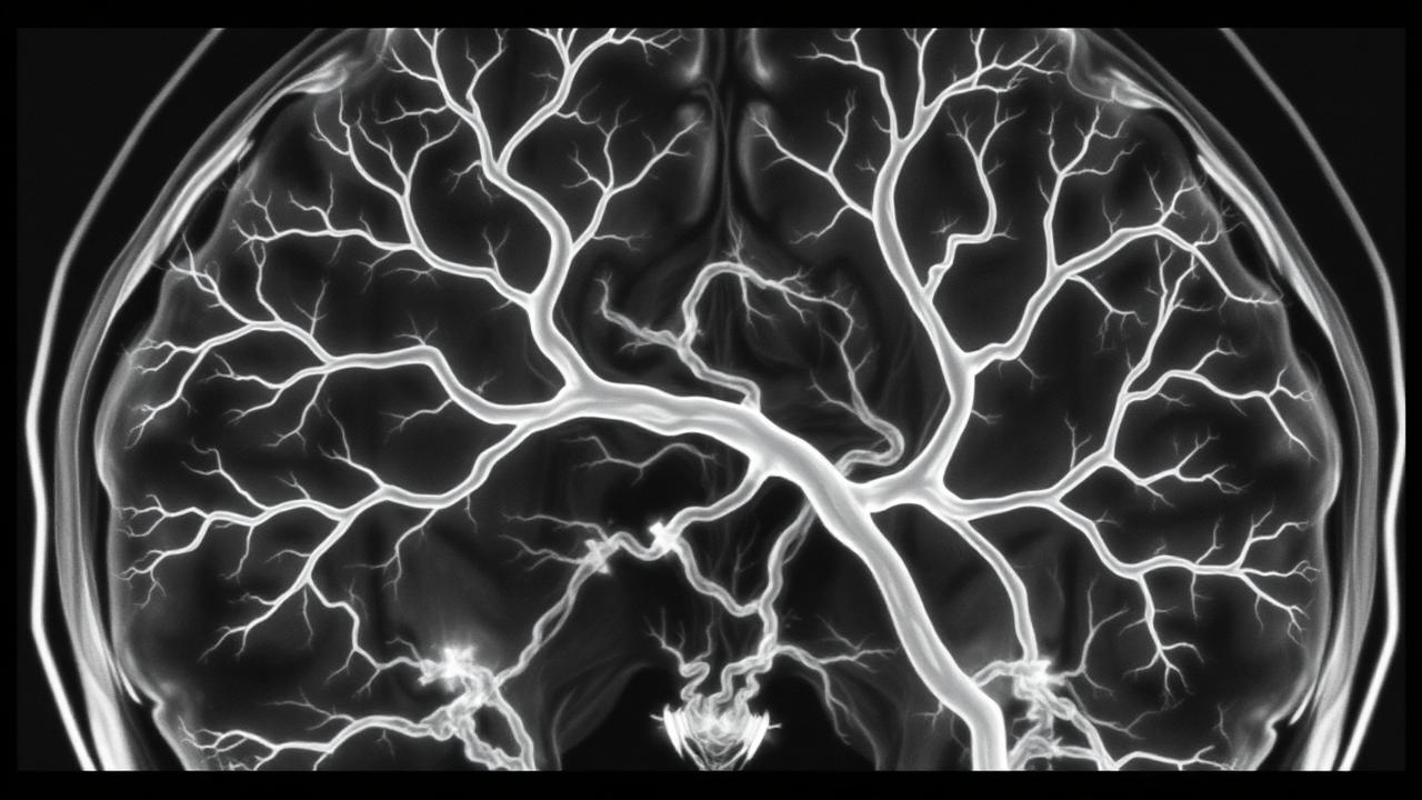
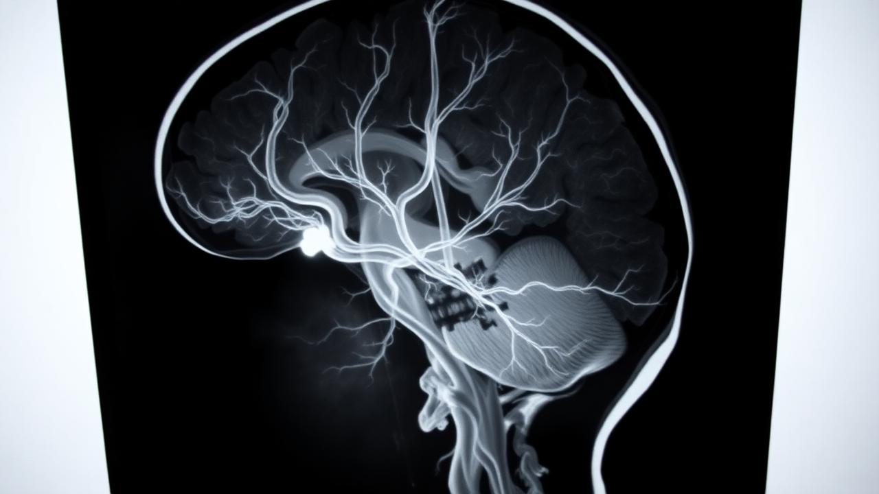
Case 01
↓ Mechanical thrombectomy
Illustrates how angiography may appear before and after clot retrieval in an eligible stroke treatment.
These are representative educational illustrations, not an identifiable patient's records, not clinical evidence of an expected result, and not a prediction of your outcome. Results and recovery vary by condition, procedure, and patient.
Recovery
Recovery is different for every procedure and patient. These stages explain what is usually planned, not how quickly any individual will recover. Your treating team's instructions take priority.
Before discharge
The treating team checks the access site, symptoms, medicines, and whether observation or a longer stay is needed.
Early recovery
Walking, lifting, wound care, and return to work depend on the procedure and your health.
Follow-up
The timing and type of follow-up are chosen for the condition and treatment performed.
Ongoing care
Some patients need long-term medicines, wound care, rehabilitation, or specialist follow-up.
If symptoms change
Follow the discharge instructions and seek urgent help for the warning signs your treating team explains.
Physician
Dr. Mandeep Sagar is a Vascular and Neuro Interventional Radiologist. The work spans the full vascular system — from cerebral aneurysms to below-knee revascularisation — performed through pinpoint access, under real-time imaging.
Verified credentials, fellowships, hospital affiliations and publications will be listed here once supplied. No claims are made on this page without source.
Specialty
Vascular & Neuro Interventional Radiology
Modalities
Fluoroscopy · CT · MR · Ultrasound guidance
Scope
Brain · Carotid · Aorta · Peripheral · Venous
Setting
Hybrid Cath Lab
On Practice
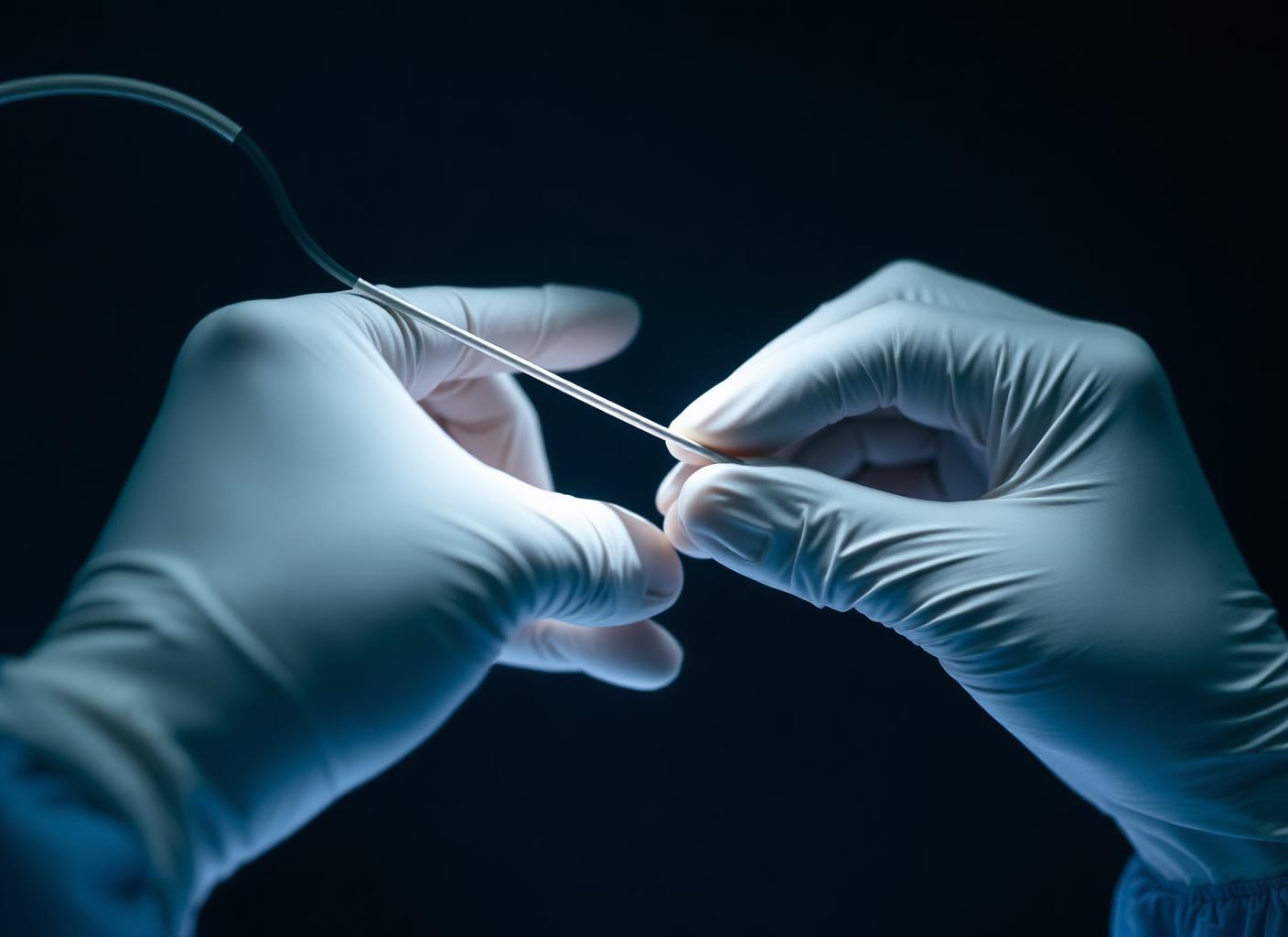
"The disease is approached where it lives — through the vessel itself."
Patient journey
Every decision begins with understanding the disease. Imaging guides the diagnosis, treatment is planned around the individual patient, and follow-up continues long after the procedure is complete.
Stage 01
Symptoms heard. History reviewed. The first decision is whether intervention is needed at all.
Stage 02
Doppler. CT angiography. MR angiography. The disease is seen before it is touched.
Stage 03
The plan is shared in plain language. Options weighed. Questions answered.
Stage 04
Every option is put on the table — including doing nothing. Risk, benefit and timing are discussed until the choice is yours.
Stage 05
Performed in a hybrid cath lab. Real-time imaging guides every millimetre.
Stage 06
Short stay. Same-day or next-day discharge for most procedures.
Stage 07
Imaging review and long-term care to keep what was restored.
Patient stories
Patient stories are published here only in the patient's own words and only with written consent. Nothing on this page is written on a patient's behalf, and no story is edited to change its meaning.
Currently
No stories are published yet. Until consented accounts are available, this section stays empty rather than filled with words no patient said.
Inside the work
Support
Understanding vascular and neuro interventional treatments.
Conditions
Explore conditions affecting the blood vessels of the body, and understand the image guided treatments available for selected cases.
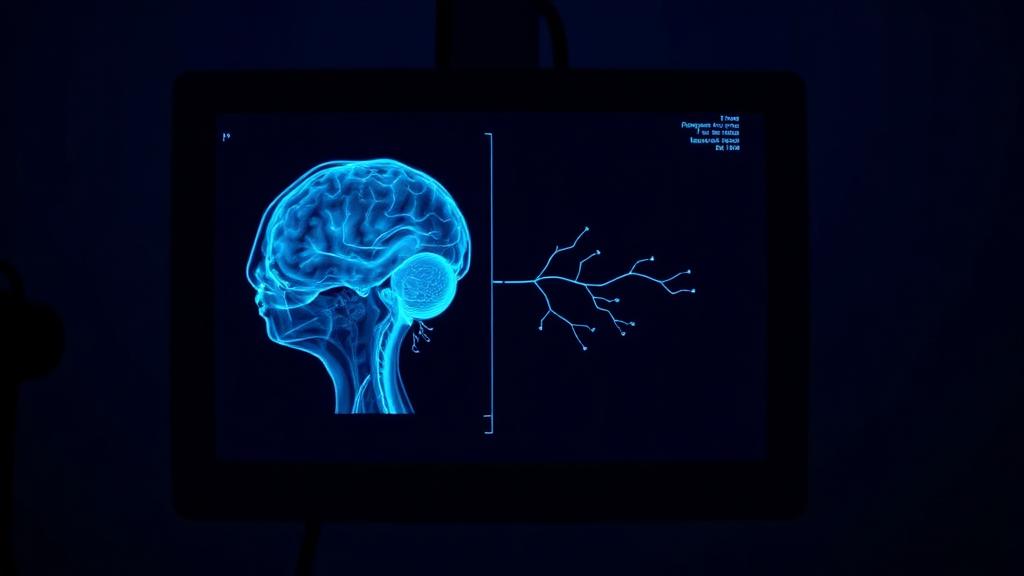
Sudden weakness, facial droop or loss of speech
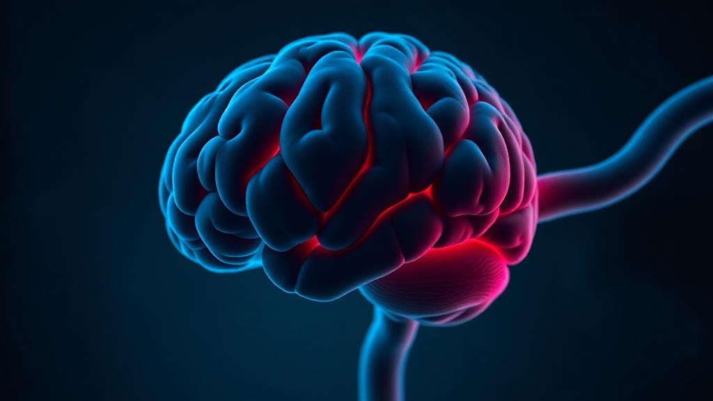
A weakened, ballooned area of a brain blood vessel
An abnormal connection between arteries and veins
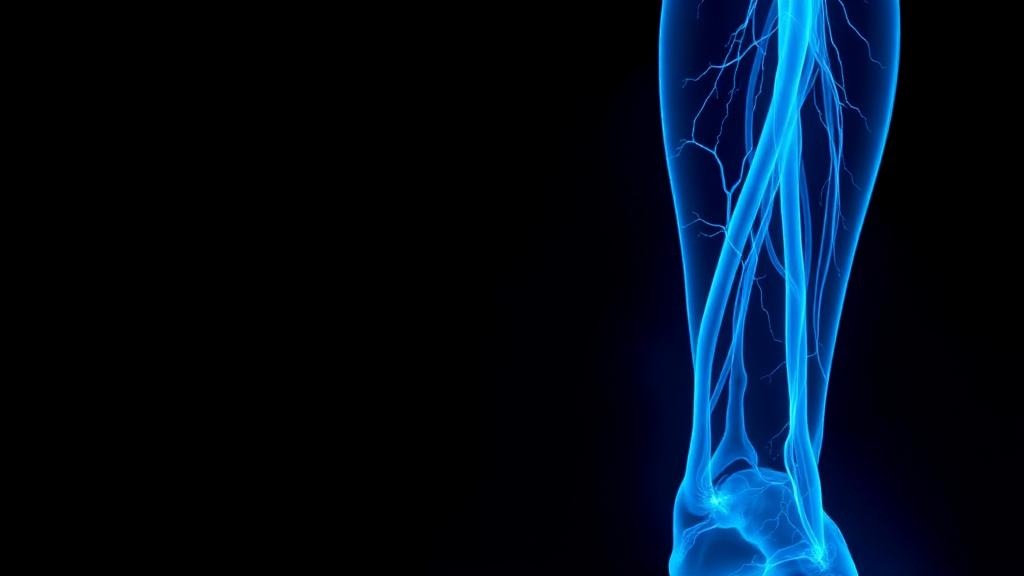
Leg pain on walking, cold feet, wounds that do not heal
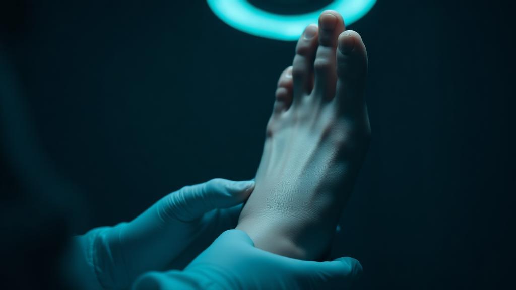
Diabetes-related foot wounds and ulcers
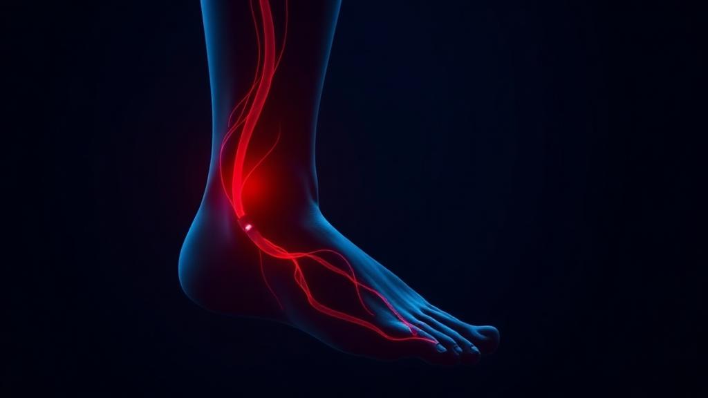
Blackened toes or dead tissue in the foot
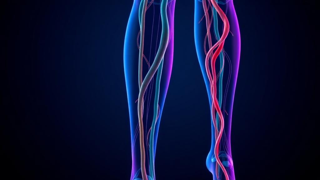
Bulging leg veins, aching legs, ankle ulcers
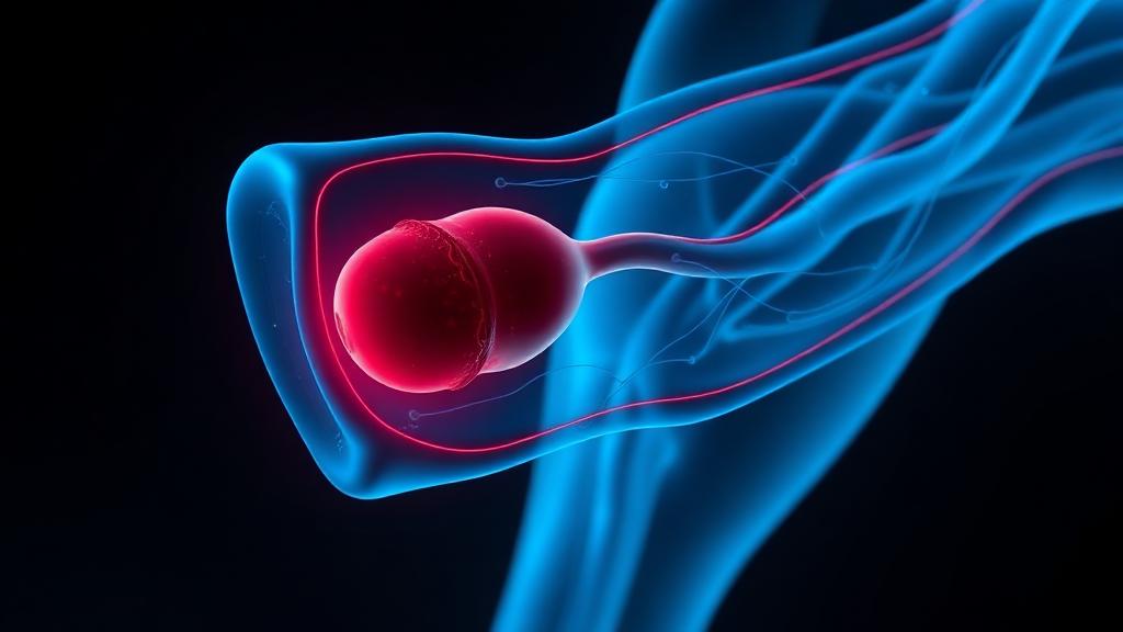
A blood clot in a deep leg vein
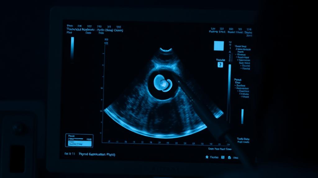
A lump or swelling in the front of the neck
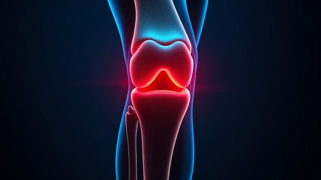
Persistent knee pain and stiffness
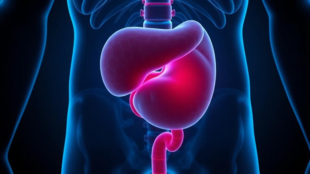
Weak urine flow and frequent night-time urination
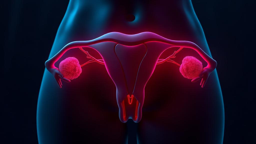
Heavy periods, pelvic pressure and pain
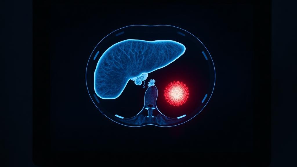
Liver cancer (HCC) and benign liver lesions
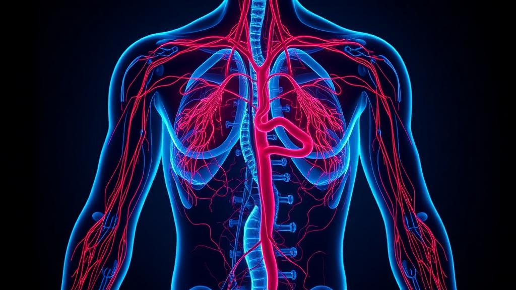
A symptom pattern, not a diagnosis on its own
Consultation
Whether you're seeking a diagnosis, a second opinion or treatment options, the first step is understanding the condition. Share your reports, speak with the team or schedule a consultation.
Book consultation
Reserve a slot with Dr. Sagar
Direct message to the practice
Call Dr. Sagar
+91 63663 30505
Patient coordinator
For relatives arranging treatment
Upload reports
Send scans and reports on WhatsApp
Reports can be shared directly on WhatsApp — PDFs, CD images, angiography stills or photographs of a wound.
Routine contact only. For an emergency, call 112 or go immediately to the nearest emergency department; do not wait for a reply here.

